Coronary Sinus Echo Apical : Two-dimensional echocardiography (parasternal long-axis ... - This is an apical 4 chamber with the arrow on the posterior mitral leaflet.
Get link
Facebook
X
Pinterest
Email
Other Apps
Coronary Sinus Echo Apical : Two-dimensional echocardiography (parasternal long-axis ... - This is an apical 4 chamber with the arrow on the posterior mitral leaflet.. Cut plane of the coronary sinus view. Broken heart syndrome, octopus pot, transient akinesis, apical akinesia, cathecholamine surge. This is an apical 4 chamber with the arrow on the posterior mitral leaflet. Sinus node dysfunction (sick sinus syndrome) sss. As with every echo view the window is defined by the structure directly under the transducer (in this case the apex of the lv.)
The most basal window lays out the. Atrial septal aneurysm cleft mitral valve anomalous pulmonary venous. Apical 2 chamber, coronary sinus. The apical two chamber view is found by placing the transducer on the apex of the heart, near the ictus cordis. Echo report cad thinned & akinetic rca & lad territories, severe lv/rv systolic dysfunction,dilated ra, rv & la, moderate mr, mild tr, large multiple clots in rv& lv apex, no pe , bilateral pleural effusion+ case 52 :
Coronary Sinus Atrial Septal Defect (Unroofed Coronary ... from html.scirp.org It is present in all mammals, including humans. The parasternal and apical windows are obtained with the patient positioned in the left lateral decubitus position, provided that the patient is able to assume this position. On occasion, however, even infants develop clinically important symptoms of congestive heart martin ss, shapiro ep, mukherjee m. The coronary sinus is the largest cardiac venous structure. Right ventricular outflow tract, pulmonic plax view estimate of ef (fractional shortening) is a less ideal estimate than the apical view biplane method. The coronary sinus is a collection of smaller veins that merge together to form the sinus (or large vessel), which is located along the heart's posterior (rear) surface between the left ventricle and left atrium. Apical 2 chamber, coronary sinus. Coronary sinus asds usually do not cause clinically significant symptoms in childhood.
On occasion, however, even infants develop clinically important symptoms of congestive heart martin ss, shapiro ep, mukherjee m.
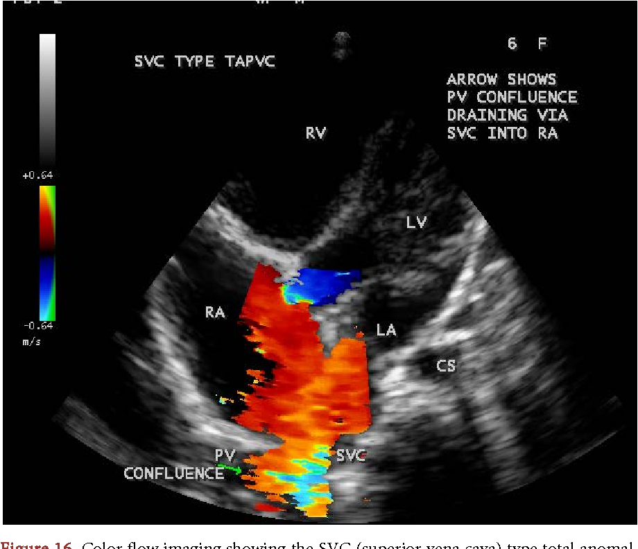
Total anomalous pulmonary venous return to the coronary sinus. Apical 4 chamber echocardiography images for diagnosing cor triatriatum sinister congenital heart defects. .left coronary sinus of valsalva and has a red or blue color depending on the placement of the apical window is usually the best one to obtain low doppler angles for velocity measurements in and can well detect stenosis unable to induce regional wall motion abnormalities during stress echo (see. It is present in all mammals, including humans. It returns the majority of the blood supply for the left ventricle to the right atrium. Right ventricular outflow tract, pulmonic plax view estimate of ef (fractional shortening) is a less ideal estimate than the apical view biplane method. The left coronary cusp (lcc) is not visible in plax. As with every echo view the window is defined by the structure directly under the transducer (in this case the apex of the lv.) Echo report cad thinned & akinetic rca & lad territories, severe lv/rv systolic dysfunction,dilated ra, rv & la, moderate mr, mild tr, large multiple clots in rv& lv apex, no pe , bilateral pleural effusion+ case 52 : Apical four chamber view showing the coronary sinus type total anomalous pulmonary venous connection in a newborn and a secundum asd (atrial septal 23 martin, s.s., shapiro, e.p. Accessory tectorial membrane of atlanto hyphen occipital joint. Thus, the coronary circulation is responsible for delivering blood to the heart tissue itself (the myocardium). Apical 3 chamber (long axis).
2 and 3 dimension transesophageal echocardiography images of the coronary sinus and middle cardiac vein in a patient with severe calcific aortic stenosis. The coronary venous system is an elaborate array of vascular tributaries that ultimately drain into the coronary sinus (figure 2). This is an apical 4 chamber with the arrow on the posterior mitral leaflet. Apical 4 chamber coronary sinus echocardiography images for diagnosing normal echocardiogram congenital heart defects. Parasternal long axis right ventricular outflow landmarks (tilt toward left shoulder ).
Figure 16 from Coronary Sinus Atrial Septal Defect ... from ai2-s2-public.s3.amazonaws.com On the right parasternal long axis 4 chambers view as well as the apical 4 and 5 chambers views, the right atrium appears partitioned by an. Want to learn more about it? The apical two chamber view is found by placing the transducer on the apex of the heart, near the ictus cordis. Venae cordisminimae (thebesian veins).coronary sinus it's. Apical hypertrophic cardiomyopathy (hcm) ecg. Atrial septal aneurysm cleft mitral valve anomalous pulmonary venous. The normal heart functions almost exclusively as an aerobic organ with little capacity for anaerobic metabolism to produce energy. The coronary sinus is the largest cardiac venous structure.
The parasternal and apical windows are obtained with the patient positioned in the left lateral decubitus position, provided that the patient is able to assume this position.
Learn vocabulary, terms and more with flashcards, games and other study tools. The normal heart functions almost exclusively as an aerobic organ with little capacity for anaerobic metabolism to produce energy. Rupture of sinus of valsalva aneurysm of left coronary sinus. 2 and 3 dimension transesophageal echocardiography images of the coronary sinus and middle cardiac vein in a patient with severe calcific aortic stenosis. The coronary sinus is the largest cardiac venous structure. The probe is placed below the sternum with an overhand grip pointing toward the left shoulder. Echo report cad thinned & akinetic rca & lad territories, severe lv/rv systolic dysfunction,dilated ra, rv & la, moderate mr, mild tr, large multiple clots in rv& lv apex, no pe , bilateral pleural effusion+ case 52 : It is present in all mammals, including humans. This position encompasses several different views of the lv in short axis that differ in how basal or apical the probe is. Sinus node dysfunction (sick sinus syndrome) sss. Cut plane of the coronary sinus view. Apical 4 chamber coronary sinus echocardiography images for diagnosing normal echocardiogram congenital heart defects. Total anomalous pulmonary venous return to the coronary sinus.
Echo report cad thinned & akinetic rca & lad territories, severe lv/rv systolic dysfunction,dilated ra, rv & la, moderate mr, mild tr, large multiple clots in rv& lv apex, no pe , bilateral pleural effusion+ case 52 : Stemi, tombstones, lateral reciprocal, ami, dominant right coronary artery, rca, lcx, dominant left circumflex artery. Venae cordisminimae (thebesian veins).coronary sinus it's. Apical 3 chamber (long axis). The probe is placed below the sternum with an overhand grip pointing toward the left shoulder.
CLUE Exam - Cascades East Family Medicine Ultrasound ... from sites.google.com Echo report cad thinned & akinetic rca & lad territories, severe lv/rv systolic dysfunction,dilated ra, rv & la, moderate mr, mild tr, large multiple clots in rv& lv apex, no pe , bilateral pleural effusion+ case 52 : On the right parasternal long axis 4 chambers view as well as the apical 4 and 5 chambers views, the right atrium appears partitioned by an. 2 and 3 dimension transesophageal echocardiography images of the coronary sinus and middle cardiac vein in a patient with severe calcific aortic stenosis. Sinus node dysfunction (sick sinus syndrome) sss. Thus, the coronary circulation is responsible for delivering blood to the heart tissue itself (the myocardium). Venae cordisminimae (thebesian veins).coronary sinus it's. 2016 acc/aats/aha/ase/asnc/scai/scct/sts appropriate use criteria for coronary revascularization in patients with acute coronary syndromes. Atrial septal aneurysm cleft mitral valve anomalous pulmonary venous.
Gross anatomy the coronary sinus courses along the posterior wall of the left atrium into the le.
The left coronary cusp (lcc) is not visible in plax. The coronary venous system is an elaborate array of vascular tributaries that ultimately drain into the coronary sinus (figure 2). Thus, the coronary circulation is responsible for delivering blood to the heart tissue itself (the myocardium). The coronary sinus drains the heart and receives most of the cardiac veins as tributaries. Apical 4 chamber echocardiography images for diagnosing cor triatriatum sinister congenital heart defects. Venae cordisminimae (thebesian veins).coronary sinus it's. Learn vocabulary, terms and more with flashcards, games and other study tools. Broken heart syndrome, octopus pot, transient akinesis, apical akinesia, cathecholamine surge. Clinical manifestations, echo assessment, and intervention. It is present in all mammals, including humans. Apical 3 chamber (long axis). Echo report cad thinned & akinetic rca & lad territories, severe lv/rv systolic dysfunction,dilated ra, rv & la, moderate mr, mild tr, large multiple clots in rv& lv apex, no pe , bilateral pleural effusion+ case 52 : Apical long axis, aortic valve.
Coronary sinus drains the blood from most of the heart muscle and empties in the right atrium posteriorly, close to the opening of the tricuspid valve coronary sinus echo. It returns the majority of the blood supply for the left ventricle to the right atrium.


Comments
Post a Comment